
Right after breast surgery, attention naturally focuses first on the overall change in size and volume;
however, as time passes and swelling subsides, many patients begin to pay close attention to their cleavage.
"Why don't they come together in the center?"
"There seems to be too much space between my breasts."
Quite a few patients share these exact concerns.
When you understand the principles behind how cleavage is formed beforehand,
you can set far more realistic and satisfying standards
rather than relying on vague expectations when planning your surgery.
Why is it that, even after breast surgery, cleavage might not appear quite as expected?
1. Does the implant determine your cleavage?
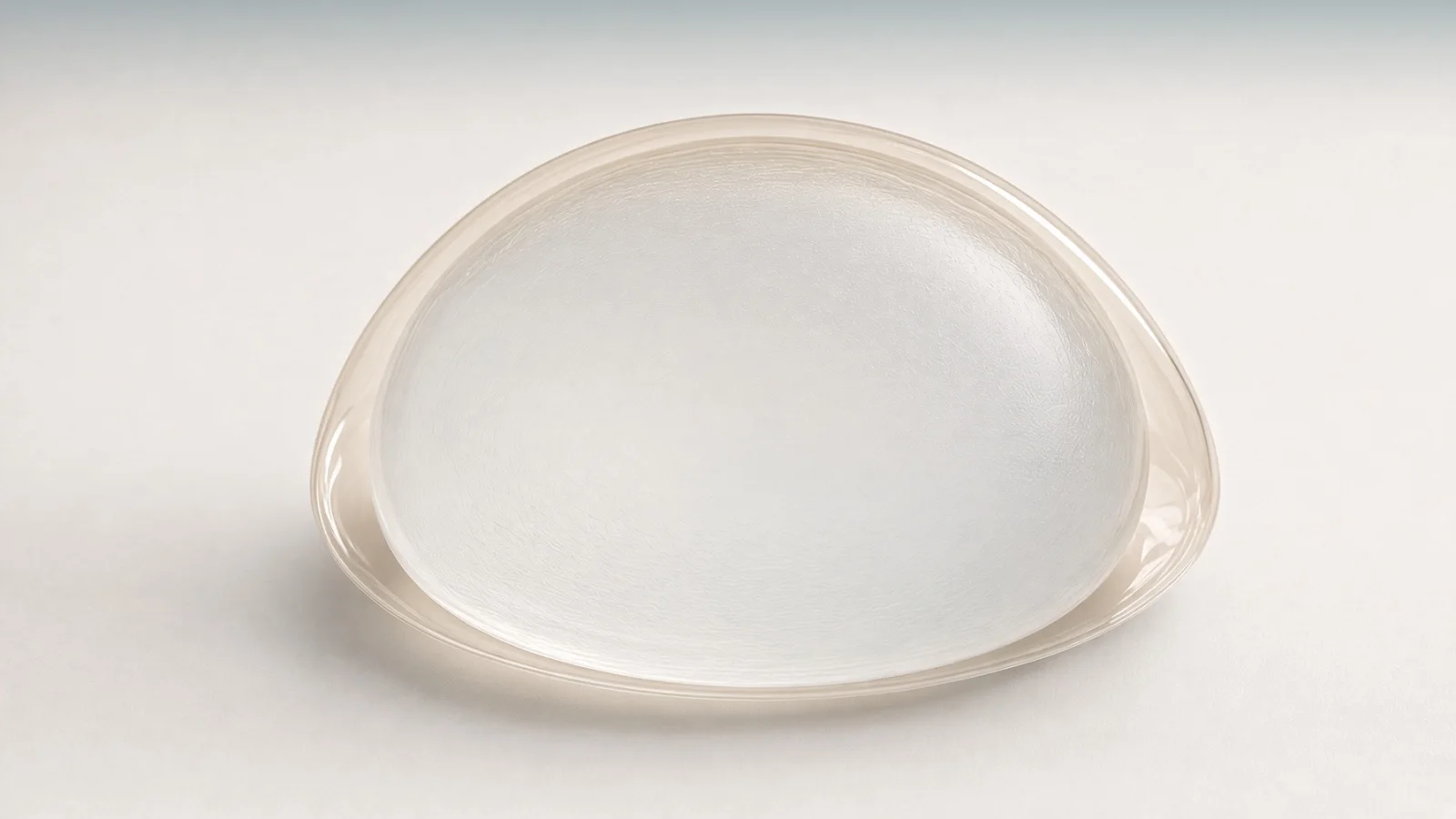
Many people anticipate that inserting voluminous implants
will naturally create deep, defined cleavage.
However, what truly determines the shape and depth of your cleavage
is not the implant itself, but the condition of your body's soft tissue covering it.
During pre-operative examinations, there are three primary anatomical factors I assess most thoroughly:
First, skin elasticity and tissue envelope flexibility.
If the skin has sufficient laxity and stretches smoothly and comfortably,
a natural medial gathering effect can be beautifully achieved once the implants are in place.
Conversely, if the skin is firm and tight,
even with the same implant size, the degree to which the breasts can gather inward will inevitably be restricted by skin tension.
Second, the tissue thickness along the inner breast.
When there is an adequate layer of subcutaneous fat and glandular tissue over the center of the chest (the sternal area),
it creates a gentle contour over the bone, providing a smooth, natural cleavage line.
However, if a patient has a leaner frame with very thin soft tissue medially,
the implant margins can become more noticeable,
often making the space between both breasts appear disconnected.
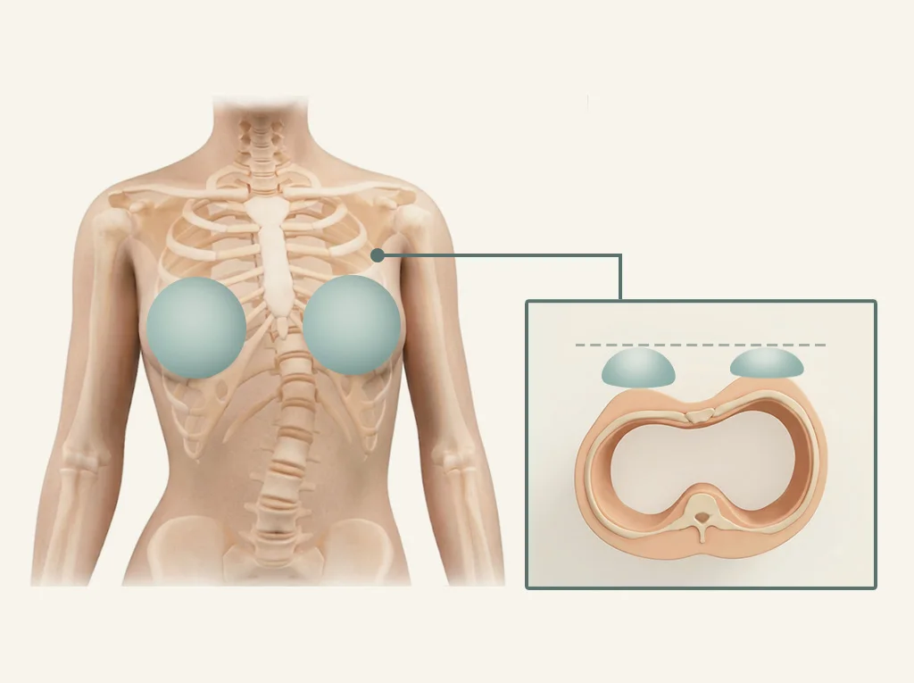
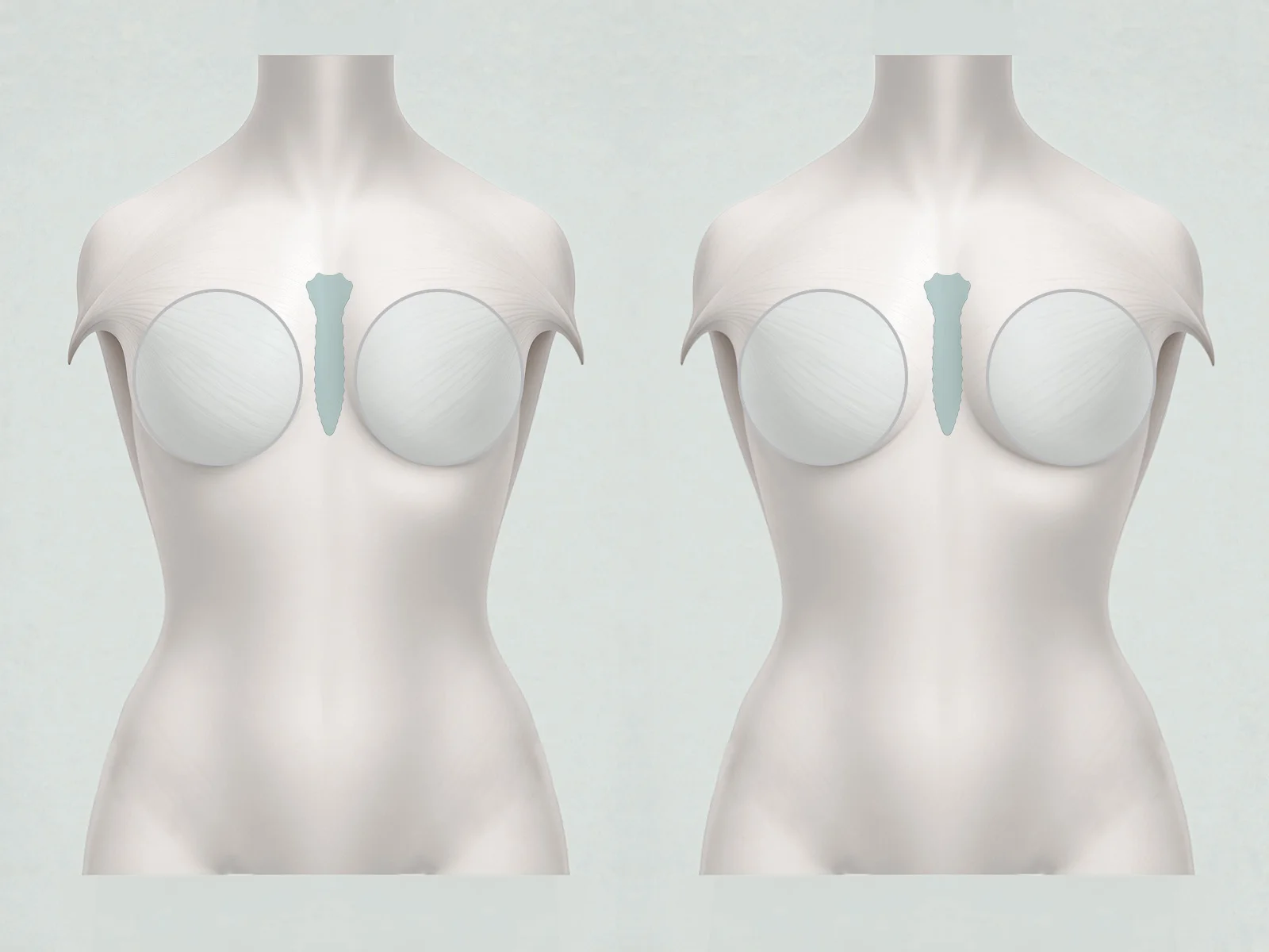
Third, the interpectoral distance.
The anatomical origin where the left and right pectoralis major muscles attach relative to the central sternum (breastbone) varies from person to person.
Particularly when performing surgery using the Dual Plane technique—where the implant is placed beneath the muscle—
if the muscles are anatomically set far apart,
there is a physical limit to how far inward the implants can be placed without excessive tension.
Ultimately, cleavage is not something newly created solely out of an implant;
it is a matter of how naturally it can be expressed over your inherent anatomical framework.
2. How much can be adjusted through implant positioning alone?
Naturally, the pocket position where the implant settles during surgery is the fundamental starting point.
If the implants are placed too far outward, the central cleavage area will appear hollow;
conversely, if they are aggressively pushed too far inward,
the overall breast shape may look unnatural, or the risk of complications such as 'symmastia'—
where the breasts bridge across the center—substantially increases.
Therefore, taking the nipple position as the central landmark,
we must first establish the most ideal and stable pocket for the implant.
Even with such precise positioning, however,
patients who inherently have thin medial soft tissue
may still feel that the gap between their breasts looks somewhat empty.
How, then, can this insufficient medial tissue thickness be addressed?
3. Can surplus body fat be used to fill the cleavage area?
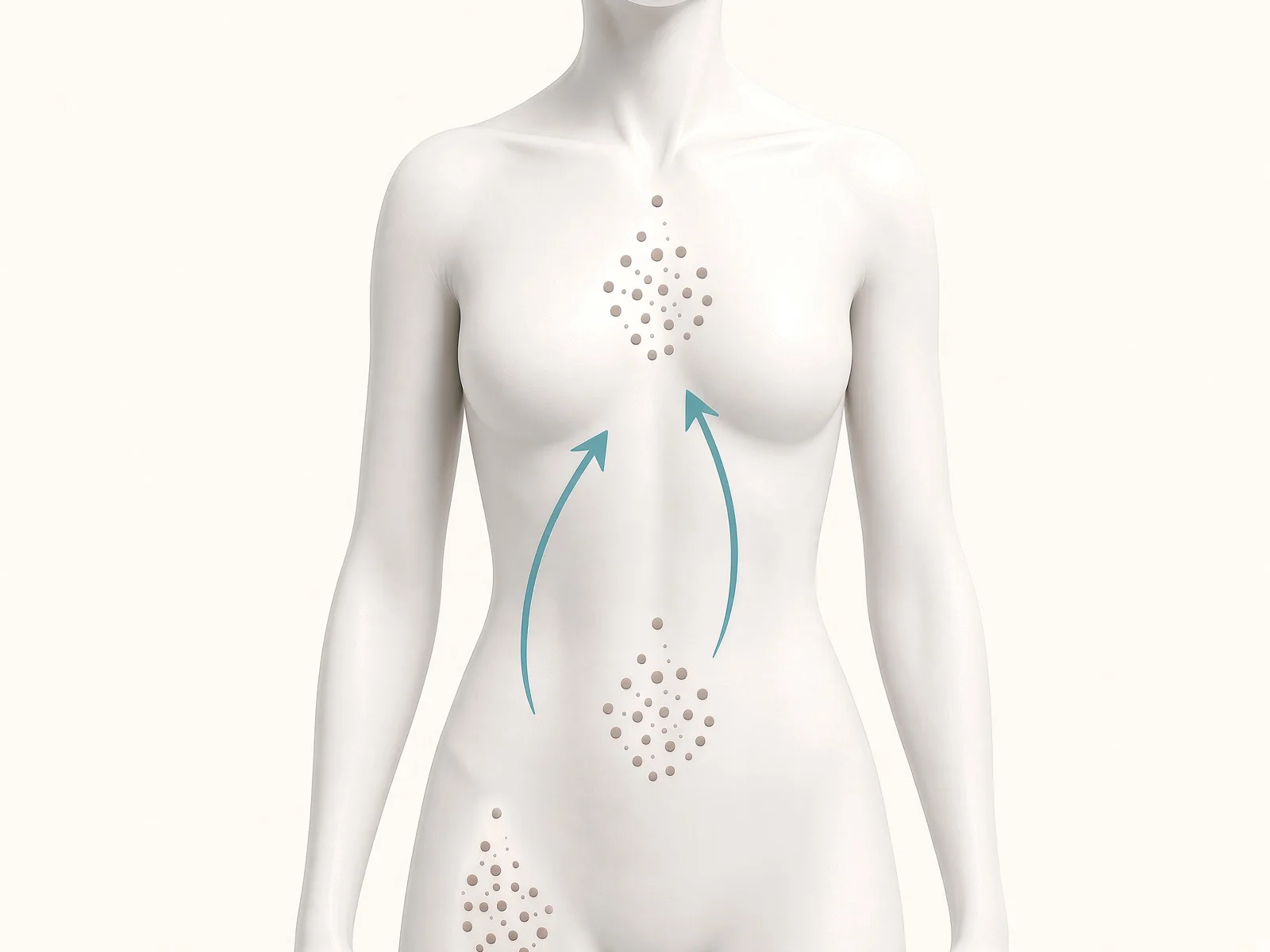
Autologous fat grafting can be used to supplement the lacking thickness along the inner breast.
This method involves harvesting unwanted fat from areas of concern such as the abdomen, flanks, or thighs,
purifying it, and meticulously grafting it into the thinner areas around the cleavage line.
By thickening the soft tissue layer covering the implant, the characteristic round contours of the implant edge soften,
resulting in a far more natural transition from the chest wall into the breast.
The key objective here is not an artificially pinched, overly tight cleavage,
but rather a soft, continuous, and comfortable line that prevents the intermammary space from appearing hollow.
If a patient is generally very lean and lacks sufficient harvestable fat,
combining finely particulated acellular dermal matrix (Particulated ADM) with fat can also be considered.
ADM is a biomaterial originally used to support and protect implants,
and it helps reinforce tissue thickness in areas where fat volume alone may not be quite enough.
In other words, we tailor our approach to each patient's physique: standalone fat grafting when sufficient fat is available,
or a combined approach with ADM when fat is limited.
4. How much of the grafted fat survives?
An essential fact to understand when planning fat grafting
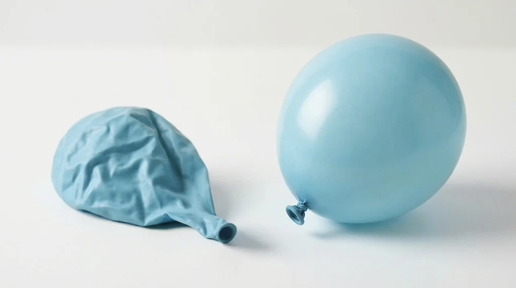
is that grafted fat does not retain 100% of its initial volume; a certain portion is naturally absorbed by the body.
For transplanted fat cells to survive, microvessels from surrounding tissues
must grow into the graft to supply adequate oxygen and nutrients.
Much like a transplanted tree needing fertile soil
to establish firm and healthy roots, the same principle applies here.

That is why in fat grafting, the focus is not simply on "how much is injected," but on "how well it survives (retention rate)."
Generally, fat graft retention rates are known to range between 50% and 70%,
and for the cleavage area, we carefully design the plan taking into account tissue characteristics, graft depth, and absorption rates associated with the surgical technique.
To support an optimal survival environment for the fat cells before new blood vessels fully revascularize the graft,
hyperbaric oxygen therapy (HBOT) can be utilized as a supportive adjunct.
Breathing high-concentration oxygen inside a pressurized chamber increases the amount of dissolved oxygen in the blood,
a mechanism that supports tissue oxygenation and promotes neovascularization in the grafted area.
While HBOT alone does not dictate the entire surgical outcome,
it serves as a valuable component of a structured aftercare regimen designed to optimize graft survival after fat transfer.
Cleavage After Breast Surgery: Key Points to Remember
Key Determinants of Cleavage
Rather than implant size or type alone, skin elasticity, medial tissue thickness, and interpectoral distance define the anatomical potential of your cleavage.
A Stepwise Approach
After precisely establishing the position of the implants,
if the medial tissue is thin, autologous fat grafting (combined with ADM if indicated) gently softens and refines the margins.
Planning for Retention
Because a portion of the grafted fat is naturally absorbed,
it is advisable to plan not only the surgical procedure itself, but also comprehensive aftercare that supports an optimal environment for graft survival.
Pre-Operative Consultation Checklist
- Does the surgeon directly evaluate your skin elasticity, medial tissue thickness, and interpectoral distance to explain realistic cleavage outcomes?
- Does your body type call for supplemental fat grafting to refine the cleavage, and if so, from which donor site will it be harvested?
- If you have a very lean frame with limited donor fat, what alternative solutions are presented?
- Is there a structured recovery and aftercare program in place to help optimize graft retention following fat transfer?
A specialist dedicated to breast surgery should always transparently explain a patient's unique anatomical conditions and boundaries
before making unconditional promises of an ideal cleavage.
Related procedure